
This new Lancet Digital Health 09/04/2026 paper titled “Digital pathology, image analysis, and artificial intelligence in liver disease” is a broad review of how digital pathology and AI could improve the analysis of liver tissue.
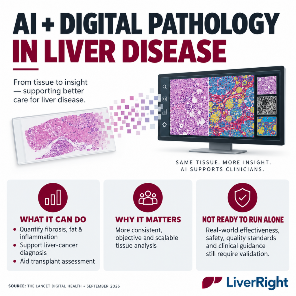
The main points:
- Traditional pathology relies on a specialist visually examining tissue under a microscope. Digital pathology converts the entire glass slide into a high-resolution image that can be viewed remotely and analyzed computationally.
- AI can measure features such as fibrosis, steatosis, inflammation and cellular abnormalities more quantitatively and consistently than conventional visual scoring alone.
- Promising applications include:
- Diagnosing and classifying primary liver cancers and metastases
- Quantifying chronic liver-disease severity
- Supporting transplant assessment, including rejection and graft quality
- Enabling remote consultations, research, education and second opinions
- The technology could reduce pathologist-to-pathologist variation, relieve laboratory workforce pressure and produce more standardized measurements for clinical trials and treatment monitoring.
- However, major obstacles remain: expensive scanners and data storage, inconsistent slide preparation and staining, interoperability problems, limited access in lower-resource settings, algorithm bias, uncertain accountability and insufficient real-world validation.
- The authors emphasize that AI should support—not replace—the liver pathologist. Algorithms developed at one hospital may not perform equally well when used with different scanners, stains or patient populations.
The most important caveat: this is a review, not a clinical trial. It does not demonstrate that AI pathology currently improves patient survival, treatment outcomes or healthcare costs. The authors conclude that real-world effectiveness, safety and implementation still require proper evaluation.
For LiverRight, the strategic implication is that digital pathology can add a highly quantitative tissue-data layer to blood tests, elastography and MRI. But it is best presented today as an emerging clinician-support capability—not yet a proven replacement for biopsy interpretation or noninvasive testing.